
Research Projects
BiyoTrap Project
The focus of the biyotrap project has been to develop a rapid detection instrument for bacteria in small concentrations.
MURI Project
This research will explore fundamental scientific issues that lead to new knowledge, understanding and technology for the prediction, diagnosis and mitigation of fuel biodeterioration and biocorrosion problems impacting US Naval operations. The project will explore to what extent the biocorrosion of carbon steel experienced in marine systems can be correlated with anaerobic fuel biodegradation, by addressing such hypotheses as: A) fuel biodeterioration and metal biocorrosion are differentially associated with the development of sulfate-reducing and/or syntrophic hydrocarbon-degrading anaerobic biofilms; B) biofuel formulations have the potential to exacerbate the baseline problem rate, depending on their susceptibility to biodegradation; and C) fuel biodegradation problems can be monitored and interrupted at sensitive stages to prevent spoilage and control biocorrosion. These hypotheses will be investigated by four research teams at four institutions, the University of Oklahoma, Colorado School of Mines, Montana State University and Oklahoma State University, in well-defined technical approaches. The MSU team's role and responsibilities include but are not limited to understanding the effect of key microbial interactions on the biocorrosion of carbon steel: This involves developing a versatile experimental platform employing a wide range of surface imaging and surface analytical techniques that will address the fundamental mechanisms of the anaerobic carbon steel biodeterioration process resulting from the anaerobic biodegradation of fuel in marine systems in the presence of sulfate and under sulfate-depleted conditions, at the nano-, micro- and macro-scales. Our results will complement and contribute to the studies carried out by our collaborators. Our team will focus on two parallel tasks designed to investigate the relationship between fuel biodegradation and biocorrosion, each with a series of specific objectives summarized under two tasks. Task 1 requires the development and employment of smart immunosurfaces (immuno-microarrays) to aid the understanding of early biodegradation/biocorrosion events at the nano- and microscales. Task 2 focuses on the comprehensive use of surface-sensitive methods such as advanced microscopy, spectroscopy , in situ microprobing and surface-sensitive analysis to evaluate the effect of biominerals and metabolites (H2S, S2-, H2) formed during the fuel degradation process on the corrosion of carbon steel at the nano-, micro- and macro-scales. The experimental design developed by the MSU team will facilitate in situ and ex situ observation and sample collection for analysis by our collaborators.
NASA Project
To image antibody antigen interaction through the use of Atomic Force Microscopy.
The DEPSCoR project has been looking at different paint samples to find correlations between AFM images and the composition of the paint. Below are some of our elemental maps showing rich locations of various chemicals in a paint surface.
DEPSCoR Project
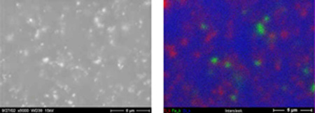
Red areas are titanium rich, green areas are iron rich, and blue are silicon rich.
To utilize Atomic Force Microscopy to image effects of marine organisms on different surfaces and the organisms themselves.
INRA
Project in conjunction with the Idaho National Environmental engineering Laboratory.
Currently the focus of this project is the characterization of known clay samples using ToF SIMS. The characterization is of the adsorption and desorption of cations as a function of laser ablation. Through the use of ToF SIMS and Origin a series of data graphs are being produced for later use.
Laser Assisted Sims (LASIMS)
Why LASIMS?
The LASIMS is being developed to meet a need of the Idaho National Engineering and
Environmental Laboratory (INEEL). Cold War era nuclear waste store at INEEL’s facility
has caused groundwater contamination.In order to analyze the contamination and characterize
the contaminants interaction with soil a new analytical technique was necessary. It
was necessary for this technique to meet two requirements: 1) Iit must generate large
fragment molecules and image their distribution on the subsurface soil contaminated
with organic and inorganic toxic materials and 2) It must increase the detection sensitivity
of the radionuclide trace elements absorbed in the subsurface soil.
Most current surface analysis techniques do not meet the objectives stated above.
Mass spectroscopy techniques offered the most promise in that not only can one have
high detection sensitivities of molecular fragments but also molecular imaging is
possible. Unfortunately the focused ion beams used for mass spec have limited probability
of desorbing large molecular fragment ions, which are very important to determine
the interaction mechanism of the toxic compounds and ions with the underlying subsurface
soil. Lasers have been used to generate large molecular fragment ions under special
sample preparation techniques such as the one used in matrix assisted laser desorption
mass spectrometry (MALDI). LASIMS takes advantage of both mass spectroscopy and laser
desorption by combining the capabilities of ToFSIMS (time of flight secondary ion
mass spectroscopy) and ToFLDMS (time of flight laser desorption mass spectroscopy).
What is LASIMS?
LASIMS proposes that SIMS and LDS can be combined to analyze a mineral surface. This technique will utilize a short laser pulse to bring the surface of the sample to just under laser desorption threshold by means of heating. A highly concentrated Ga+ beam that is well synchronized to the laser pulse will then be used to further energize the sample surface to yield molecular desorption from that surface. One problem with SIMS alone technique is the fact that information is gathered from a close proximity of the point of impact of the primary ion beam, but the laser used with the LASIMS bethod will bring to desorption threshold a large area. The result is that molecular information can now be gathered from an undamaged area in the vicinity of the point of impact of the Ga+ beam. This method has the advantage over unassisted SIMS in that the yield of chemical information from a minimal microscopic area is greatly increased.
How it works?
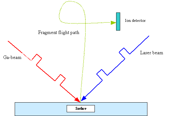
Figure 1: Schematic representation of the LASIMS method. The primary ion beam (Ga+) is preceded by a short laser pulse. (Click to enlarge)
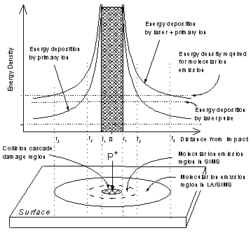
Figure 2: Schematic representation of the LASIMS method. The primary ion beam (Ga+) is preceded by a short laser pulse. (Click to enlarge)
LASIMS proposes that SIMS and LDS can be combined to analyze a mineral surface. This technique will utilize a short laser pulse to bring the surface of the sample to just under laser desorption threshold by means of heating. A highly concentrated Ga+ beam that is well synchronized to the laser pulse will then be used to further energize the sample surface to yield molecular desorption from that surface. One problem with SIMS alone technique is the fact that information is gathered from a close proximity of the point of impact of the primary ion beam, but the laser used with the LASIMS bethod will bring to desorption threshold a large area. The result is that molecular information can now be gathered from an undamaged area in the vicinity of the point of impact of the Ga+ beam. This method has the advantage over unassisted SIMS in that the yield of chemical information from a minimal microscopic area is greatly increased.
Finished Projects
MAP student program to use Atomic Force Microscopy and Scanning Electron Microscopy to measure the accuracy of an Anatech Hummer VII sputter coater and to measure the roughness of three different coatings: gold, gold/palladium, and platinum. Click for results.